Dermatome Map Upper Limb – If you have ever wondered how the human dermatome map looks, you’ve come to the right place. Before we move on to the map, let’s discuss what a dermatome is. What are the different kinds? The most important thing is what is the reason to be aware of dermatomes order to know more about your body. Read on to find out more. You may be surprised! Here are some examples of dermatomes.
Dermatomes Neurology Medbullets Step 1
What is a Dermatome?
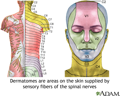
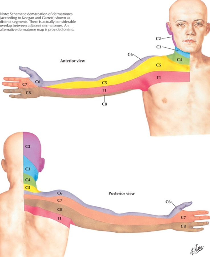
” Dermatomes” refers to the spinal cord “dermatome” refers to a tissue that is a part of the cord of the spinal. Dermatomes are important in allowing doctors to construct maps of the spinal cord that help in diagnosing. Two major maps are regarded as valid by medical professionals. These are: the Keegan and Garret map and the Foerster map. The maps were designed in the 1930s and remain frequently utilized. The trigeminal and maxillary nerve are the biggest dermatomes.
Dermatomes are skin-like areas that are attached to a particular nerve. In cases of spinal cord injury, the pain could be felt in a dermatome which is surrounded by the nerve. Similar to the pain that is caused by an outbreak of shingles can be felt in specific spinal nerves. If you suffer from pain or neurological condition involving the dermatome area, you must consult a physician.
ALSO READ:[show-list showpost=5 category=”dermatome-map” sort=sort]
What are Some Examples of Dermatomes?
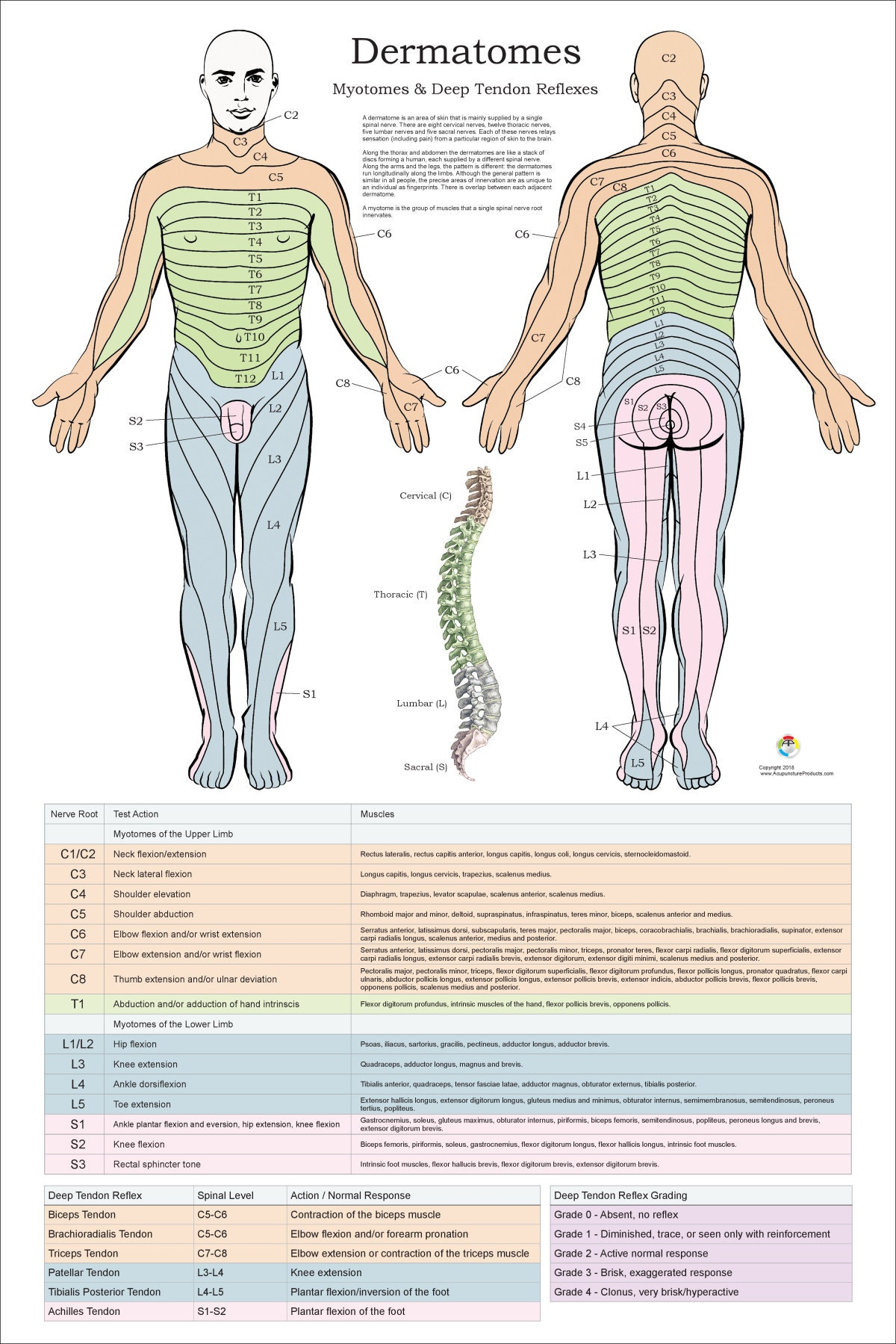
Dermatomes are a part of skin supplied by one spinal nerve. The nerves transmit motor, sensory and autonomic signals. They form part of the peripheral nerve system, which connects brain and other parts of the body. Dermatomes can suffer from a spinal injury. When one of these dermatomes gets injured, it is able to be treated easily with a local anesthetic.
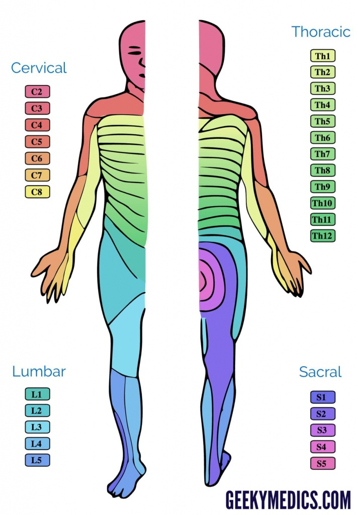
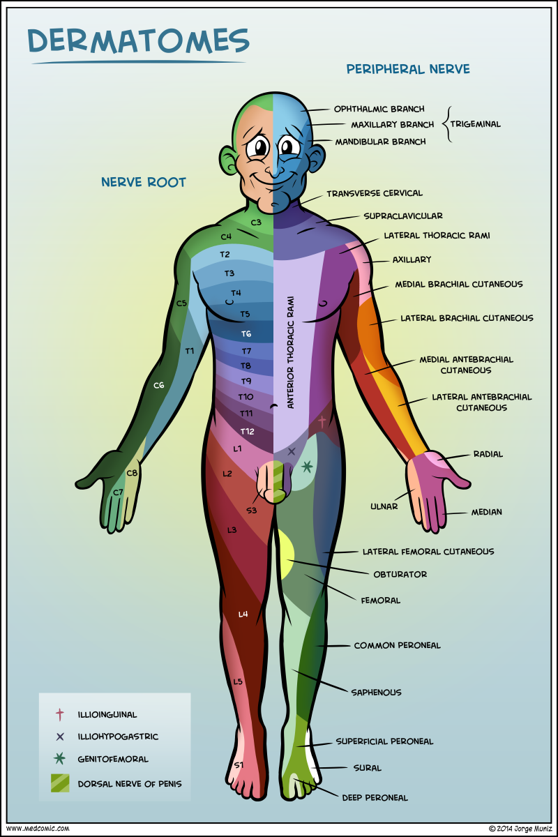
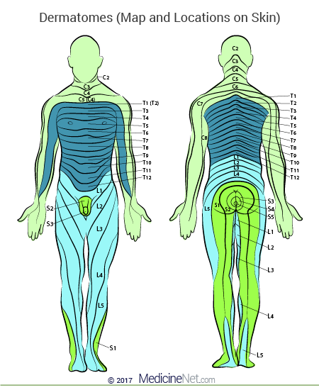
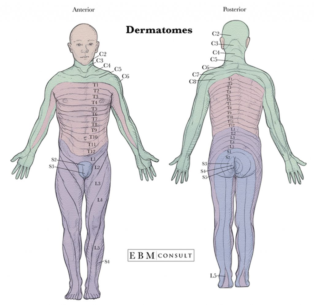
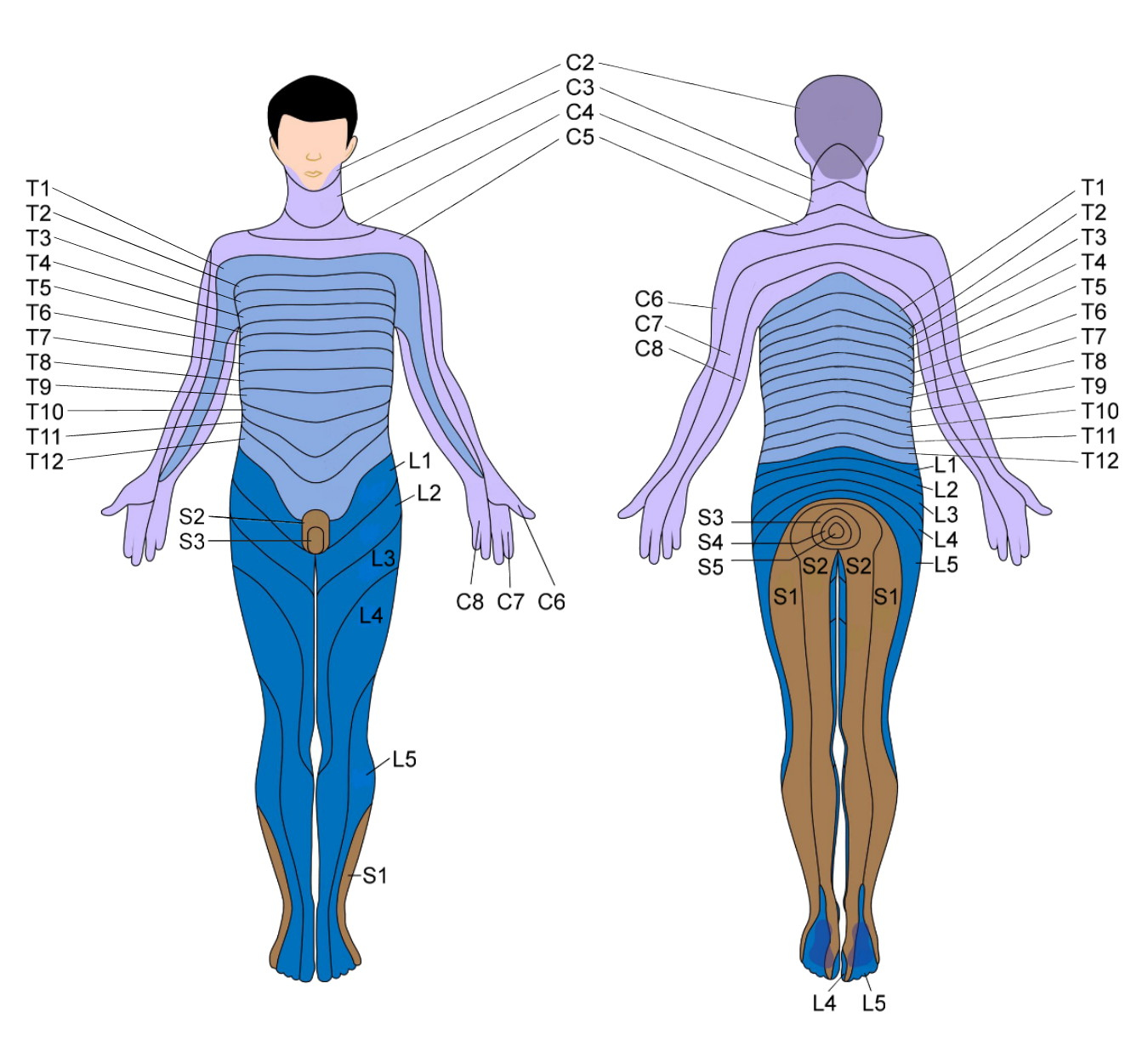
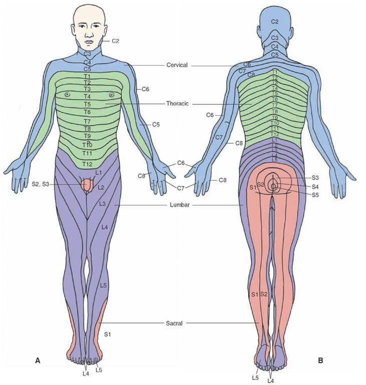
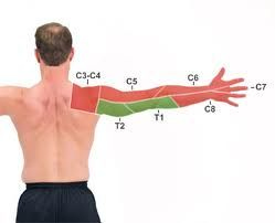
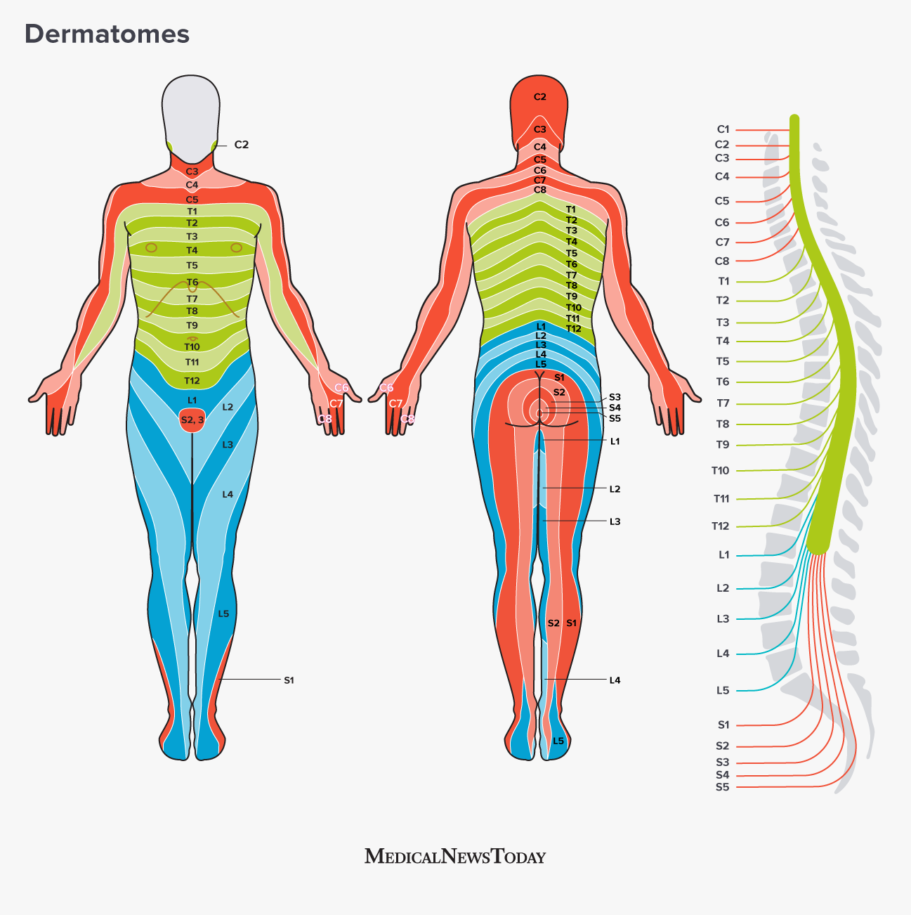
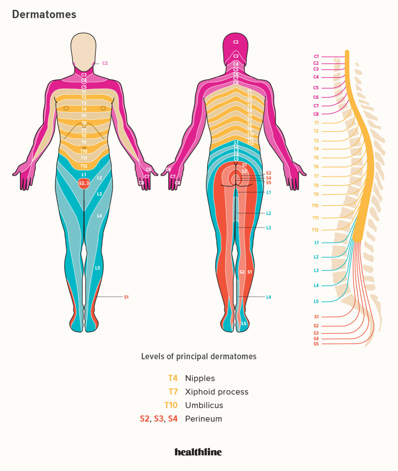
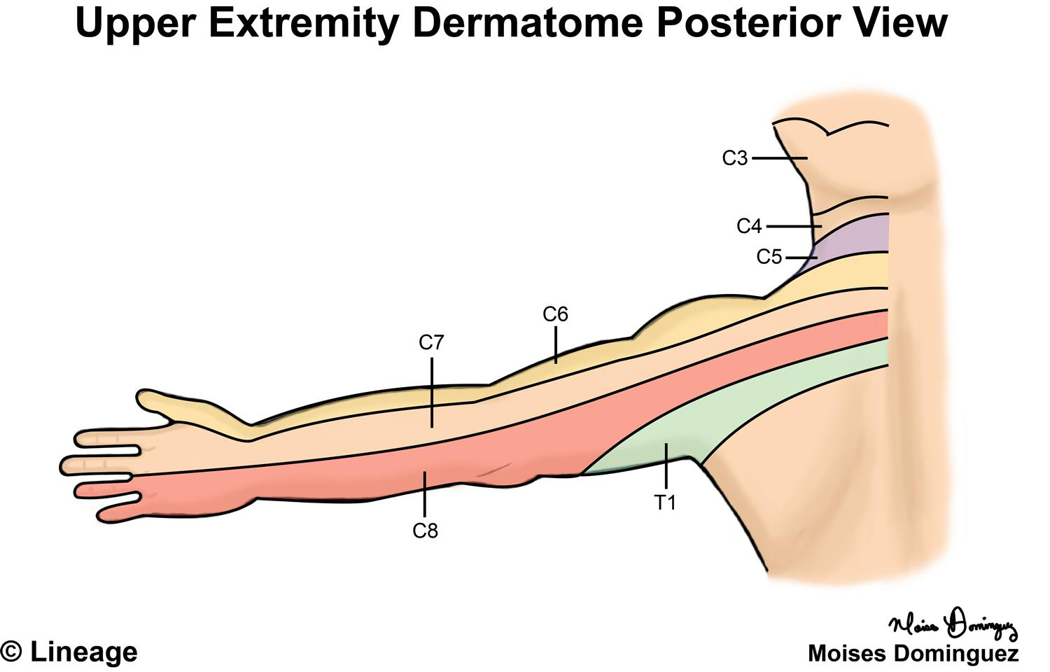
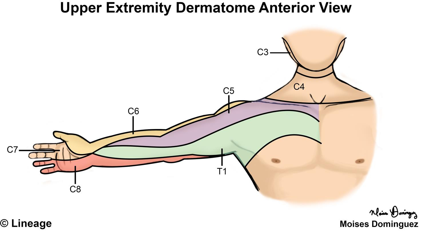
The dermatomes of the thoracic region are identified by letter-number combinations, which show the relationship between the area along with the sensor nerve that serves that region. For example C1’s spinal nerve doesn’t have a dermatome, but the other spinal nerves are identified as C1-C8 and T9 is a reference to the belly button. Dermatomes are laid horizontally on the trunk, and dermatomes located on the extremities tend to be linear.
Dermatome Map
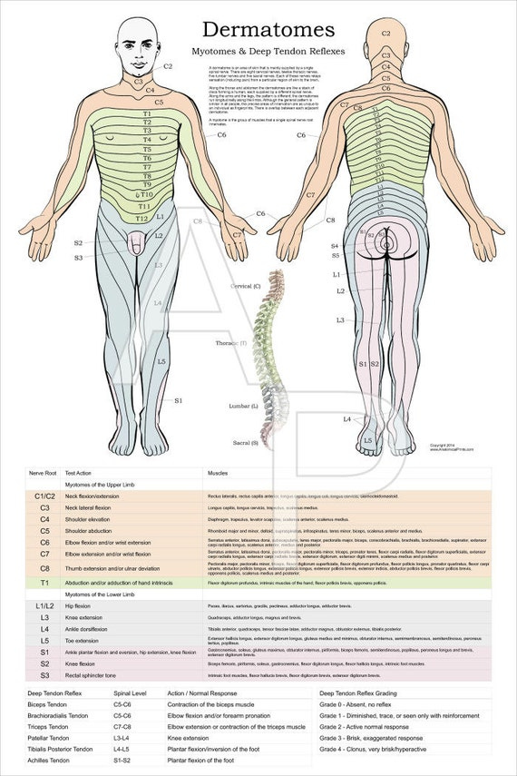
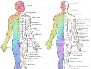
Dermatome maps are the most common element in textbooks that teach anatomy. However, the dermatome maps is not consistent both within and inter-textbook. The names are inconsistent as are some textbooks that have distinct maps on different pages. This is particularly problematic when the authors of several chapters do not agree on the selection of dermatome maps. A majority of textbooks employ the diagrams drawn by Foerster, Keegan, and Garrett however they don’t provide adequate references. Additionally, four textbooks employ maps with no citations. This includes one that uses only secondary sources.
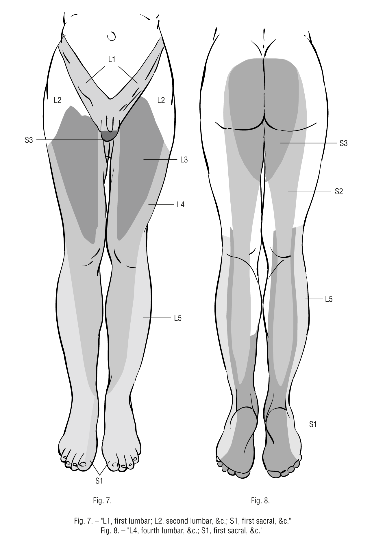
The dermatome is the area of the skin that receives sensory information from the dorsal branch of one spinal nerve. Dermatomes aren’t evenly found, but they tend to dip lower than horizontally. This is a natural variation, and some tissues are covered by more than one dermatome. Also dorsal spinal roots could contain intrathecal intersegmental connections with sensory neurons of the dorsal parts of the limbs.
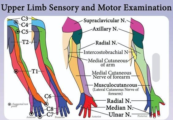
Dermatome Map Upper Limb – Dermatome Map
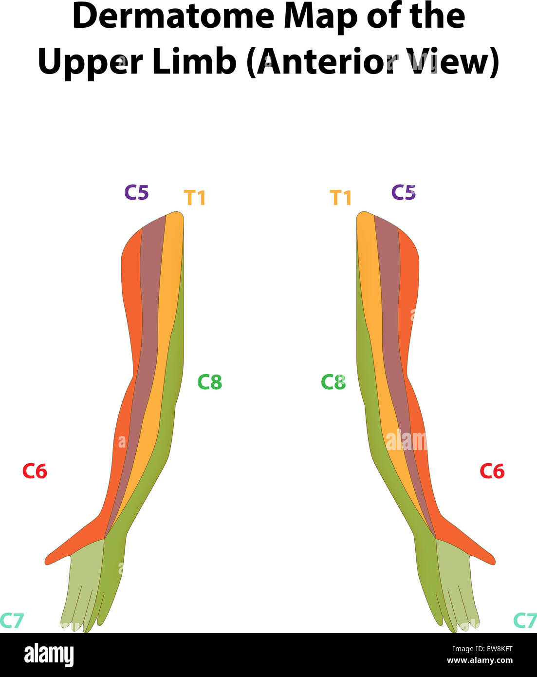
Dermatome Map Of The Upper Limb Stock Photo Alamy
Dermatomes Neurology Medbullets Step 1
dermatome Hashtag On Twitter