DERMATOMES AND MYOTOMES CHART PDF – If you have ever wondered how the human dermatome map looks, you’ve come to the right place. Before we move on to the map, let’s discuss the definition of a dermatome. What are the various kinds? And most importantly, why is it important to understand dermatomes in order to better understand how the body works. Continue reading to learn more. You might be amazed! Here are some examples of dermatomes.
Dermatomes Myotomes And DTR Poster 24 X 36 Chiropractic Etsy
What is a Dermatome?
” Dermatomes” refers to the spinal cord “dermatome” refers to a tissue that covers the spinal cord. Dermatomes can help doctors to develop images of spinal cord that can be useful in diagnosing. Two major maps are regarded as valid by medical professionals. The Keegan and Garret map and the Foerster map. These maps were made in the 1930s, and are commonly utilized. The trigeminal and maxillary nerves are the two largest dermatomes.
Dermatomes are skin areas that are attached to a specific nerve. In the case of spinal cord injuries, pain may be felt in a dermatome that is controlled by the nerve. Similar to the pain that is caused by shingles outbreaks can be felt in particular spinal nerves. If you experience a pain or neurological condition involving the dermatome, it is recommended that you visit a doctor.
ALSO READ:[show-list showpost=5 category=”dermatome-map” sort=sort]
What are Some Examples of Dermatomes?
Dermatomes are the segments of skin that is provided by one spinal nerve. These nerves carry motor, sensory, and autonomic messages. They form an element of the peripheral nervous system which connects brain and other parts of the body. A dermatome may become affected due to a spinal injury. If one of these dermatomes becomes injured, it can be treated easily with an local anesthetic.
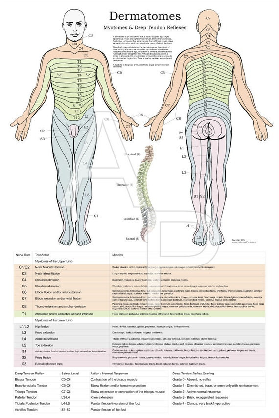
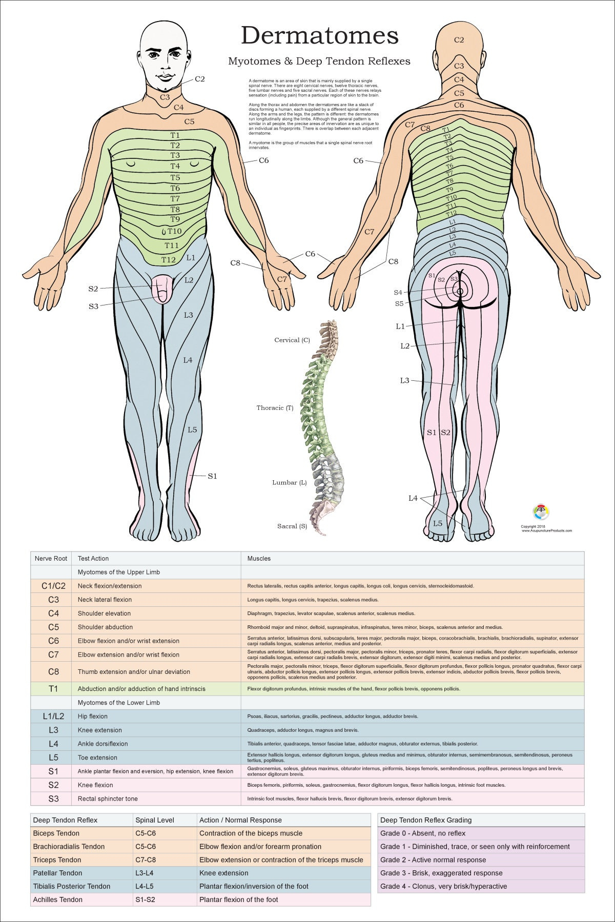
The dermatomes of the thoracic region have been labeled with letter-number combinations that show the connection between the region along with the sensor nerve that supplies the area. For instance, the C1 spinal nerve doesn’t have a dermatome, but all spinal nerves in the region are labeled as C1-C8 and T9 is a reference with the belly button. Dermatomes are layered vertically on the trunk while dermatomes on the extremities tend to be longitudinal.
Dermatome Map
Dermatome maps are the most common element in textbooks teaching anatomy. But, the map is inconsistent both intra and inter-textbook. The name is not consistent and some textbooks include distinct maps on different pages. This is particularly problematic when the authors of different chapters do not agree on the selection of dermatome map. Many textbooks use the map of Foerster, Keegan, and Garrett but do not include the proper references. Furthermore, four textbooks make use of maps with no citations, and one of them is one that cites only secondary sources.
The dermatome is the area of the skin that receives sensory information from the dorsal root of one spinal nerve. Dermatomes aren’t uniformly located, but they tend to dip lower than horizontally. This is a normal variation and some tissue types are covered with more than one. Additionally dorsal spinal roots could be anastomosed with intrathecal intersegmental sensory neurons in the dorsal parts of the limbs.
Dermatome And Myotome Map – Dermatome Map
DERMATOMES AND MYOTOMES CHART PDF