Dermatom L5 S1 The L5 S1 Spinal Motion Segment Also Called The – If you’ve ever wanted to know what the human dermatome map will look, you’re in the right spot. Before we move on to an image, it’s important to talk about what is a dermatome. What are the various kinds? And most importantly, why is it essential to understand dermatomes in order to understand how the body works. Read on to find out more. You may be surprised! Here are some examples of dermatomes.
Dermatomes Diagram Spinal Nerves And Locations
What is a Dermatome?
The term “dermatome” refers to a tissue that is a part of the spinal cord. Dermatomes play a crucial role in allowing doctors to create models of the cord, which aid in the diagnosis. Two maps are widely accepted by medical experts. There is the Keegan and Garret map and the Foerster map. These maps were made in the 1930s and are still widely used. The trigeminal nerve , as well as the maxillary nerve are the biggest dermatomes.
Dermatomes are skin areas that connect to a specific nerve bundle. In cases of spinal cord injury, pain may be felt in a dermatome that is connected to that nerve. Similarly, the pain caused by an outbreak of shingles can be felt in specific spinal nerves. If you feel nerve pain or neurological problem affecting the dermatome area, you must consult a physician.
ALSO READ:[show-list showpost=5 category=”dermatome-map” sort=sort]
What are Some Examples of Dermatomes?
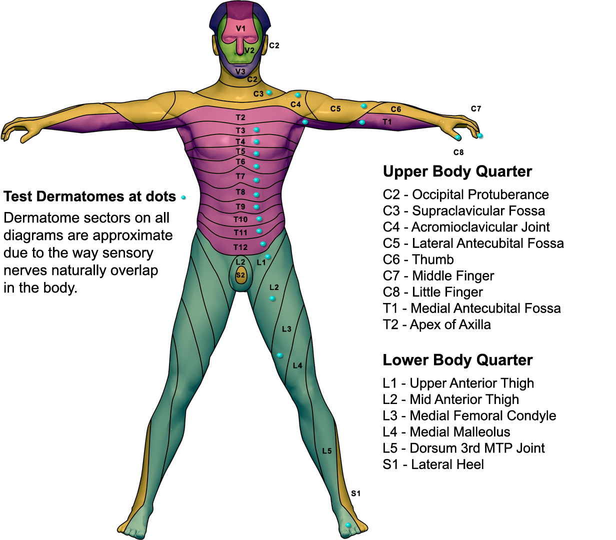
Dermatomes are the segments of skin that is provided by the spinal nerve. The nerves transmit sensory, motor as well as autonomic information. They form a part of the peripheral nervous system which connects the brain and all the body. Dermatomes can become affected due to a spinal cord injury. When one of these dermatomes is injured, it can be treated easily with the use of a local anesthetic.
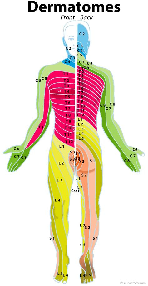
Dermatomes in the thoracic region are labeled with letters-numbers that illustrate the relationship between the area as well as the nerve which supplies that region. For instance the C1 spinal nerve doesn’t have a dermatome, but others spinal nerves have been labeled C1-C8, while T9 corresponds to the belly button. Dermatomes are layered in horizontally along the trunk, while dermatomes that are located on the extremities are generally longitudinal.
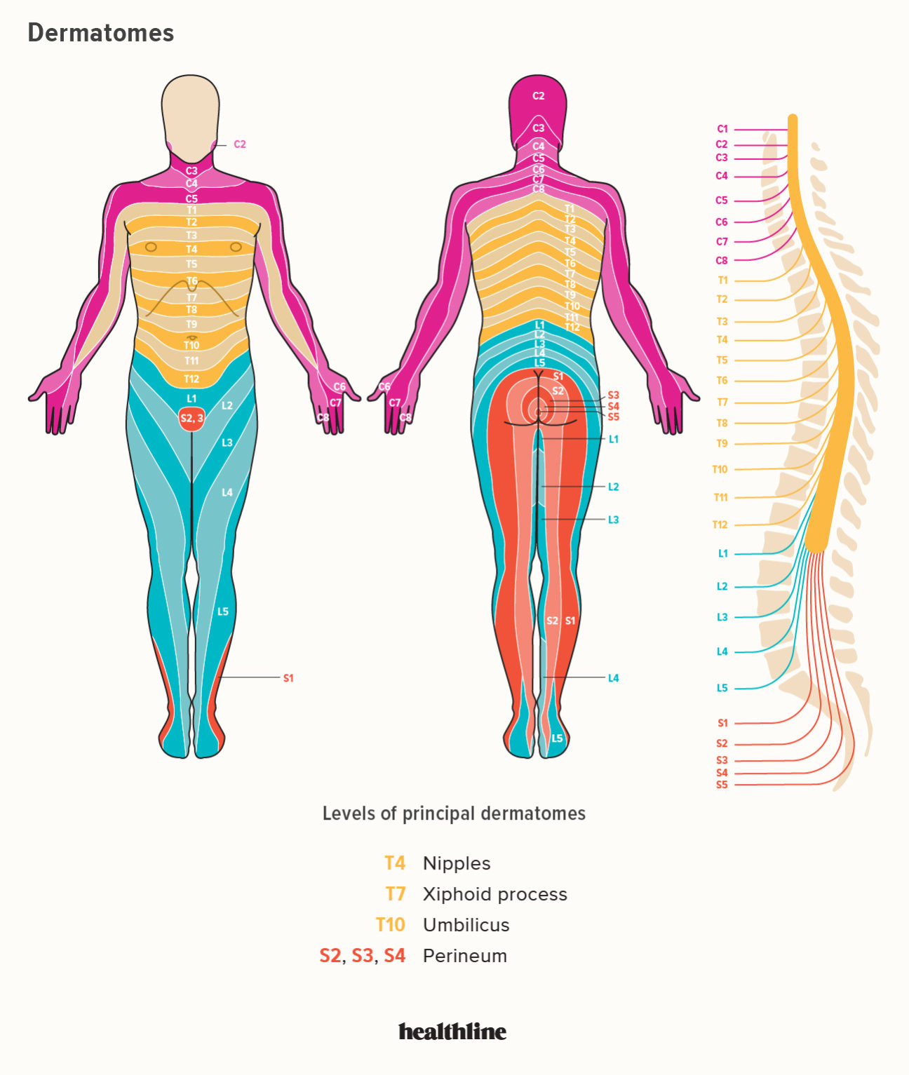
Dermatome Map
Dermatome maps are an integral part of textbooks that teach anatomy. The dermatome map is inconsistency both within and inter-textbook. The name is not consistent and certain textbooks have various maps on different pages. This can be particularly challenging when the authors of different chapters differ in their choice of dermatome maps. A majority of textbooks employ the map of Foerster, Keegan, and Garrett but don’t include appropriate references. In addition, four textbooks utilize maps that do not have citations, such as one that uses only secondary sources.
The dermatome is the area of skin that receives sensory stimulation from the dorsal root of one spinal nerve. Dermatomes aren’t evenly found, but they tend to dip less inferiorly than horizontally. This is a normal variation and certain tissues may be covered by multiple dermatomes. Also, dorsal spinal rootlets may contain intrathecal intersegmental connections with sensory neurons of the dorsal parts of the limbs.
Dermatome Cervical Map – Dermatome Map
Anterior Dermatome Map QxMD
Dermatom L5 S1 The L5 s1 Spinal Motion Segment Also Called The