
Dermatomes Definition Chart And Diagram – If you have ever wondered what the human dermatome’s map will look, you’re in the right spot. Before we get to the map, let’s look at what is a dermatome. What are the various kinds? And, most importantly, why is it essential to know about dermatomes in order to better understand the human body. Read on to find out more. You may be surprised! Here are some examples of dermatomes.
Dermatome Map Shingles Leg
What is a Dermatome?
The term “dermatome” refers to a tissue that is a part of the cord of the spinal. Dermatomes can help doctors to create maps of the spinal cord that can be useful in diagnosing. Two major maps are accepted by medical professionals. The Keegan and Garret map and the Foerster map. These maps were developed in the 1930s and are still widely used. The trigeminal nerve , as well as the maxillary nerve are the largest dermatomes.
Dermatomes are skin regions that connect to a particular nerve bundle. In cases of spinal cord injury, the pain could be felt in a dermatome which is connected to that nerve. Similarly, the pain caused by an outbreak of shingles can be felt in particular spinal nerves. If you suffer from nerve pain or neurological problem affecting the dermatome area, you must visit a doctor.
ALSO READ:[show-list showpost=5 category=”dermatome-map” sort=sort]
What are Some Examples of Dermatomes?
Dermatomes are a part of skin supplied by only one spinal nerve. These nerves carry motor, sensory as well as autonomic information. They form part of the peripheral nervous system, that connects the brain to the all the body. Dermatomes can suffer from a spinal lesion. When one of these dermatomes becomes injured, it could be treated easily with the use of a local anesthetic.
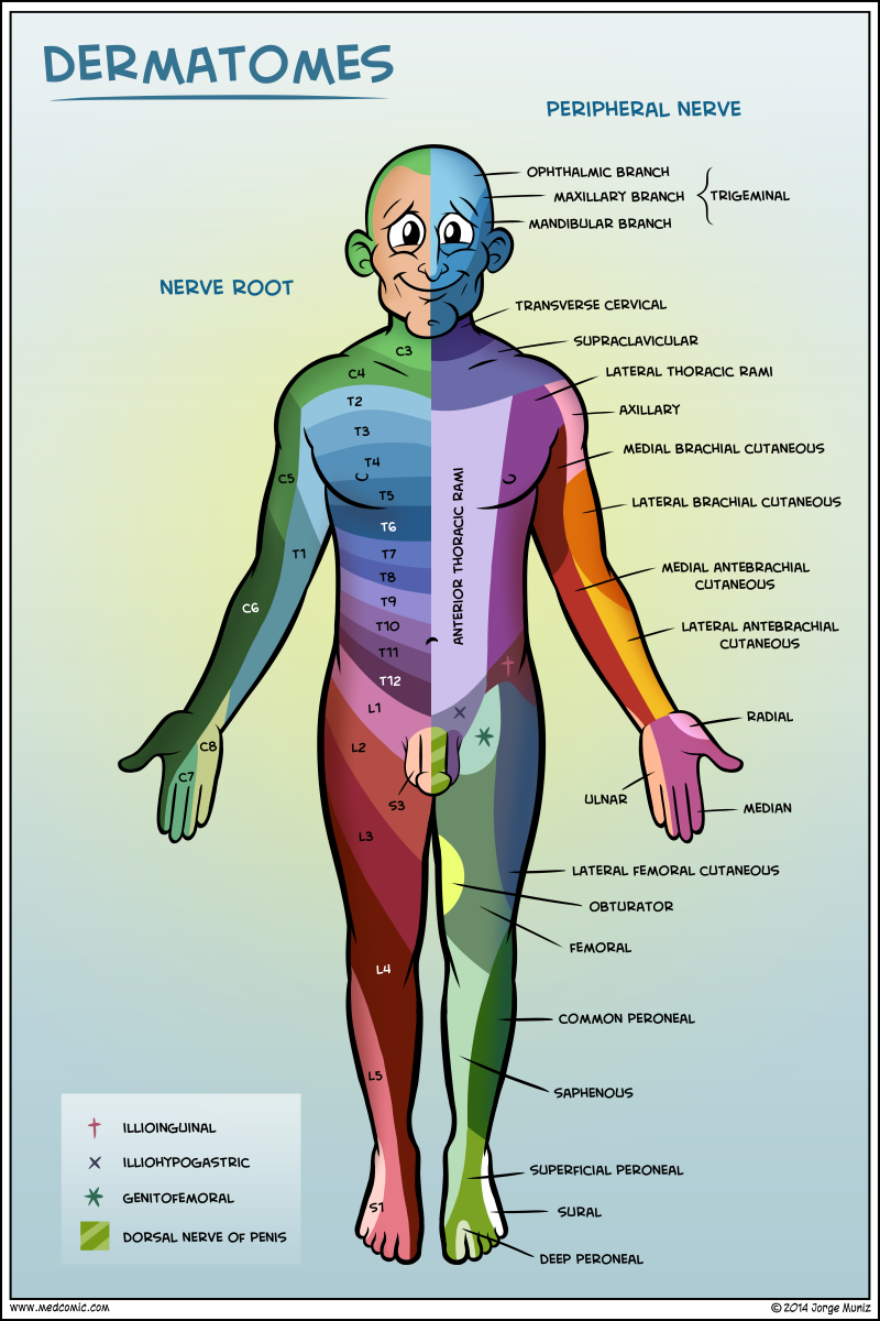
The dermatomes of the thoracic region are labeled with letters-numbers that illustrate the connection between the region along with the sensor nerve that is responsible for that area. For instance C1’s spinal nerve does not have a dematome, however others spinal nerves have been identified as C1-C8 T9, which corresponds with the belly button. Dermatomes are layered in horizontally on the trunk however, dermatomes on the extremities are typically in a longitudinal.
Dermatome Map
Dermatome maps are an integral part of textbooks that teach anatomy. The dermatome map is inconsistency both within and inter-textbook. The name is not consistent as are some textbooks that have different maps on different pages. This can be particularly challenging when the authors of different chapters disagree on the choice of dermatome maps. The majority of textbooks utilize the maps of Foerster, Keegan, and Garrett however they don’t provide proper references. Additionally, four textbooks employ maps with no citations. This includes one that uses only secondary sources.
Dermatomes are the areas of skin that receives sensory innervation from the dorsal roots of one spinal nerve. Dermatomes aren’t always evenly situated, but they tend to be more inferior than horizontally. This is a natural variation, and certain tissues have more than one. Furthermore, dorsal spinal rootlets may be anastomosed with intrathecal intersegmental sensory neurons that originate from Dorsal limbs.
Dermatome Map For Shingles – Dermatome Map
Dermatomes Definition Chart And Diagram