Dermatomes Diagram Spinal Nerves And Locations – If you have ever wondered what the human dermatome’s map appears, then you’re at the right place. Before we move on to this map, lets discuss what a dermatome actually is. What are the various kinds? Most importantly, why is it essential to learn about dermatomes in order to better understand your body. Continue reading to learn more. You might be surprised! Here are some examples of dermatomes.

Nervous System Basicmedical Key
What is a Dermatome?
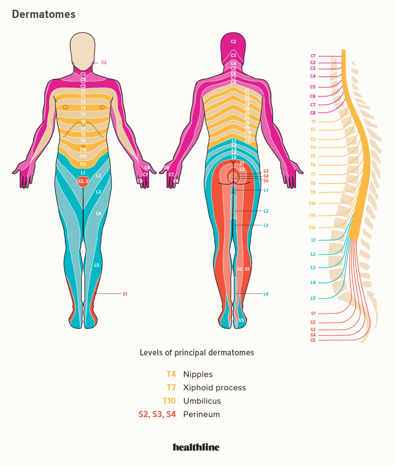
The term “dermatome” refers to a tissue that covers the cord of the spinal. Dermatomes can help doctors to develop diagrams of the spine that are useful for diagnosis. Two maps are widely accepted by medical professionals. The Keegan and Garret map and the Foerster map. These maps were created in the 1930s, and are often employed. The trigeminal nerve as well as the maxillary nerve are among the most extensive dermatomes.
Dermatomes are skin regions that are linked to a specific nerve. In the case of spinal cord injury, pain may be felt in a dermatome that is connected to that nerve. The same is true for the pain caused by shingles outbreaks can be felt in specific spinal nerves. If you suffer from discomfort or neurological issue involving the dermatome, you should see a doctor.
ALSO READ:
What are Some Examples of Dermatomes?
Dermatomes are the segments of skin that is supplied by one spinal nerve. These nerves provide sensory, motor and autonomic messages. They form an element of the peripheral nervous system which connects the brain and all the body. A dermatome may be affected by a spinal cord injury. If one of these is injured, it can be treated easily with the use of a local anesthetic.
The dermatomes of the thoracic region are labeled by letter-number combinations, which show the connection between the region in question and the sensory nerve which supplies the area. For instance, the C1 spinal nerve does not have a dematome, however the other spinal nerves are labeled C1 – C8 and T9 is a reference to belly button. Dermatomes are layered horizontally along the trunk, and dermatomes located in the extremities are usually in a longitudinal.
Dermatome Map
The dermatome map is an integral part of textbooks teaching anatomy. But, the map is inconsistent both intra and inter-textbook. The name is not consistent, and some textbooks feature distinct maps on different pages. This can be particularly challenging when the authors of different chapters disagree on the choice of dermatome maps. Most textbooks use maps of Foerster, Keegan, and Garrett however, they do not provide proper references. In addition, four textbooks utilize maps with no citations, and one of them is one that cites only secondary sources.
Dermatomes are the areas of skin that receives sensory input from the dorsal root of one spinal nerve. Dermatomes aren’t evenly located, but they tend to dip less inferiorly than horizontally. This is a natural variation and some tissues may be covered by multiple dermatomes. Also dorsal spinal nerve roots may have intrathecal intersegmental anastomoses to sensory neurons of the dorsal parts of the limbs.
Dermatome Map Nerve Paths That Shingles Follow – Dermatome Map

Pin On Inspirations

Spinal Nerves Boundless Anatomy And Physiology
Dermatomes Diagram Spinal Nerves And Locations


