Dermatome Map Shingles Leg – If you’ve ever wondered what the human dermatome’s map looks, you’ve come to the right place. Before we go to this map, lets take a look at what a dermatome actually is. What are the different types? And, most importantly, why is it essential to know about dermatomes in order to know more about the human body. Read on to find out more. You may be surprised! Here are some examples of dermatomes.
Shingles UF Health University Of Florida Health
What is a Dermatome?
“dermatome,” or “dermatome” refers to a tissue that covers the spine. Dermatomes play a crucial role in allowing doctors to develop models of the cord that help in diagnosing. Two major maps are accepted by medical experts. There is the Keegan and Garret map and the Foerster map. These maps were developed in the 1930s and are frequently utilized. The trigeminal nerve as well as the maxillary nerve are the largest dermatomes.
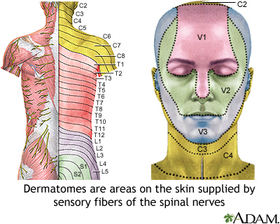
Dermatomes are skin regions that connect to a particular nerve bundle. In cases of spinal cord injury, the pain could be felt in a dermatome which is surrounded by the nerve. In the same way, the pain triggered by shingles outbreaks is felt by specific spinal nerves. If you experience a pain or neurological condition involving the dermatome region, you need to visit a doctor.
ALSO READ:[show-list showpost=5 category=”dermatome-map” sort=sort]
What are Some Examples of Dermatomes?
Dermatomes are the segments of skin that is provided by a single spinal nerve. These nerves provide sensory, motor and autonomic messages. They form an element of the peripheral nervous system, which connects brain and other parts of the body. Dermatomes can get affected because of a spinal lesion. If one of these dermatomes is injured, it can be easily treated with a local anesthetic.
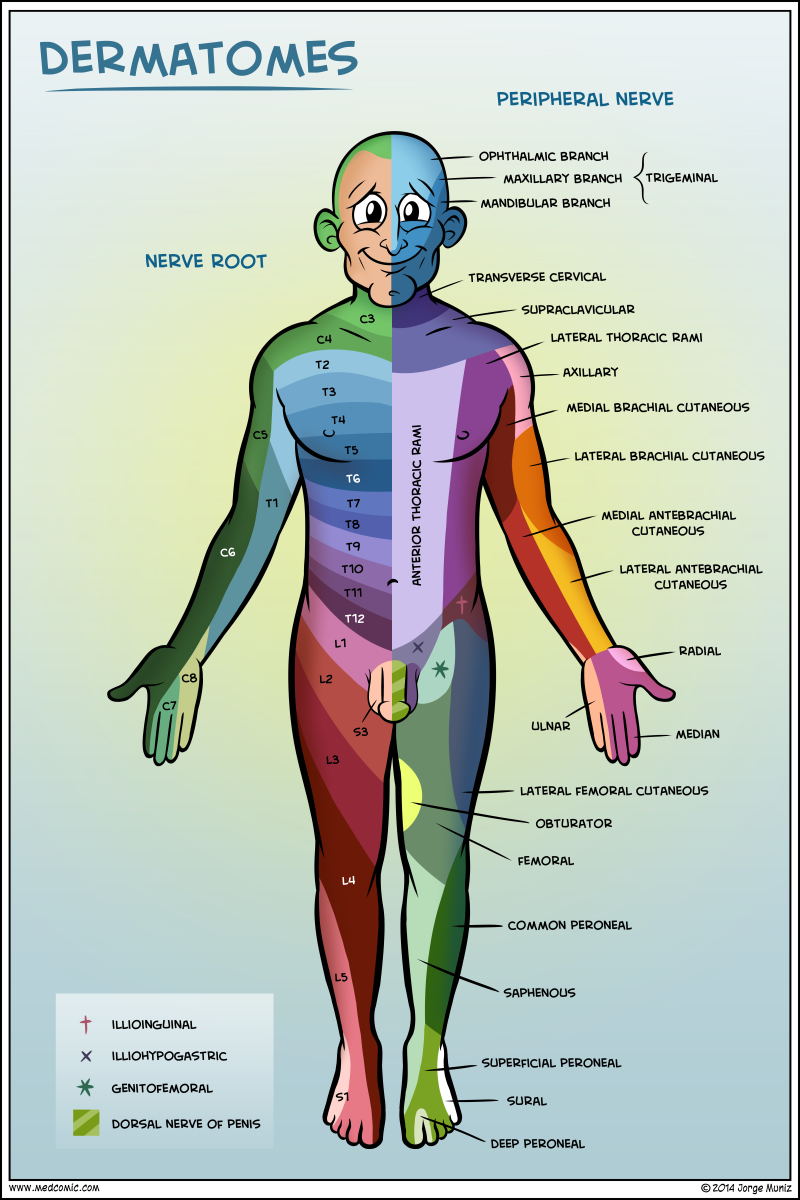
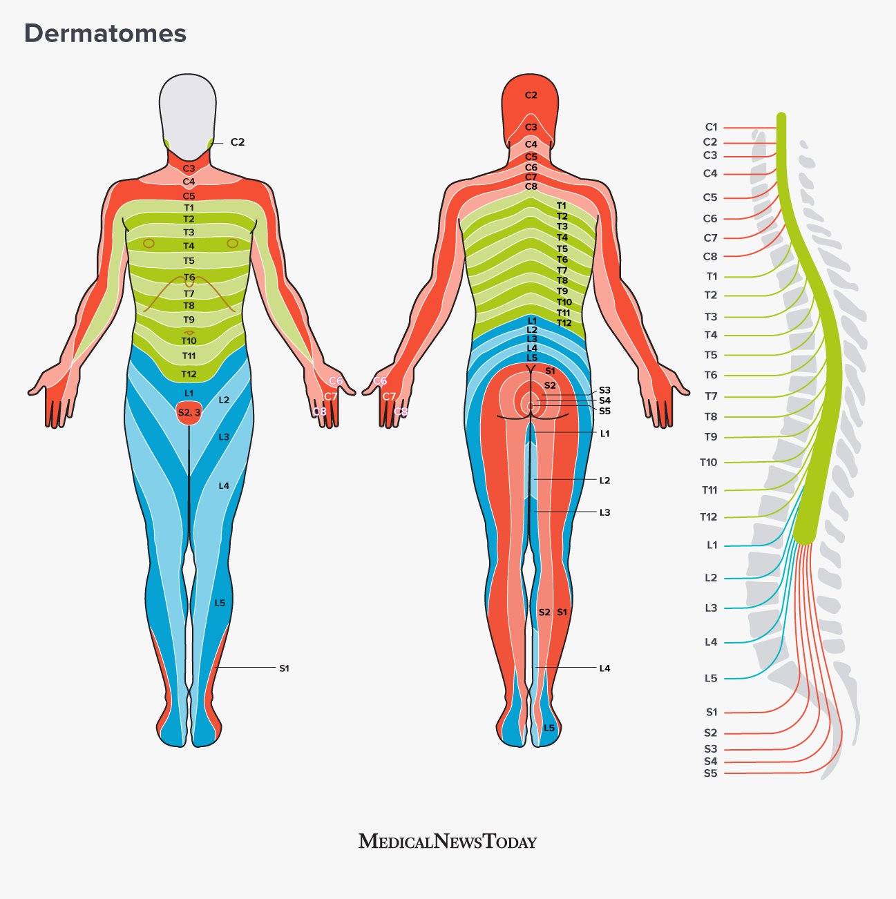
The dermatomes of the thoracic region are identified with letter-number combinations that show how the region is connected and the sensory nerve that is responsible for that region. For instance, the C1 spinal nerve doesn’t possess a dermatome, however the other spinal nerves are labeled C1 – C8 and T9 refers to the belly button. Dermatomes are laid horizontally on the trunk, however, dermatomes on the extremities tend to be long.
Dermatome Map
Dermatome maps are the most common element in textbooks that cover anatomy. But, the map is inconsistent both intra and inter-textbook. The names are inconsistent, and some textbooks feature different maps on different pages. This is particularly problematic when the authors of multiple chapters disagree on the choice of dermatome maps. A majority of textbooks employ the map of Foerster, Keegan, and Garrett but do not include appropriate references. Additionally, four textbooks employ maps that do not have citations, such as one that only cites secondary sources.
Dermatomes are the areas of skin that receives sensory stimulation from the dorsal branch of one spinal nerve. Dermatomes aren’t evenly located, but they tend to dip more inferiorly than horizontally. This is a natural variation and some tissues may be covered by multiple dermatomes. Additionally dorsal spinal nerve roots may be anastomosed with intrathecal intersegmental sensory neurons of those limbs that are dorsal.
Dermatome Map Shingles – Dermatome Map
Dermatomes Definition Chart And Diagram
Dermatome Map Shingles Leg