Dermatomes Definition Chart And Diagram – If you’ve ever wondered what the human dermatome’s map looks, you’ve come to the right place. Before we look at an image, it’s important to discuss what a dermatome actually is. What are the various types? And most importantly, what is the reason to learn about dermatomes in order to understand your body. Continue reading to learn more. You might be amazed! Here are some examples of dermatomes.
What is a Dermatome?
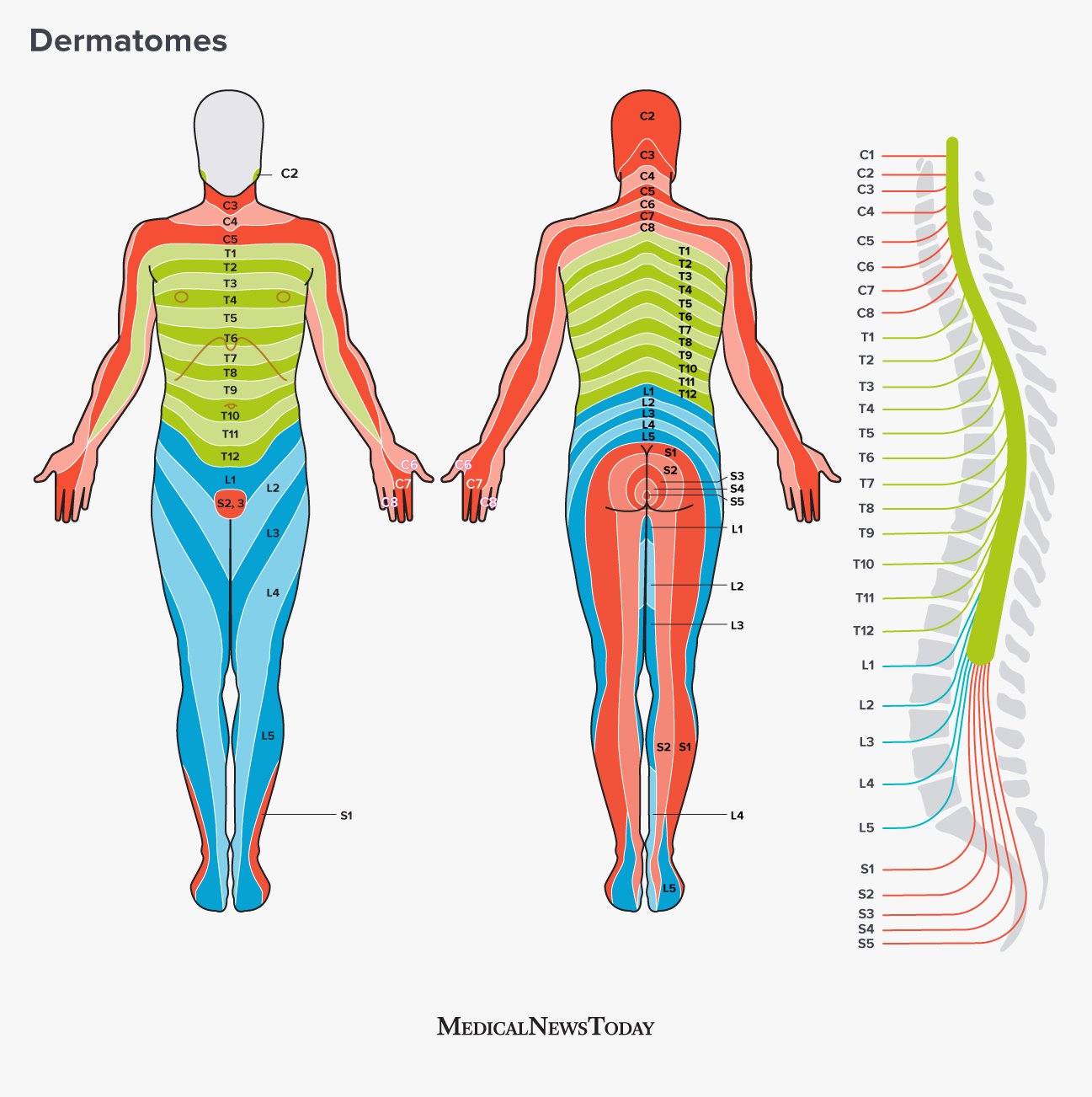
“dermatome” or “dermatome” refers to a tissue that covers your spinal cord. Dermatomes can help doctors to develop models of the cord that can be useful in diagnosing. Two major maps are accepted by medical experts. They are the Keegan and Garret map and the Foerster map. The maps were designed in the 1930s and are frequently used. The trigeminal nerve as well as the maxillary nerve are the biggest dermatomes.
Dermatomes are skin-like areas which are connected to a specific nerve bundle. In the case of spinal cord injury, the pain could be experienced in a dermatome that is innervated by that nerve. The same is true for the pain caused by an outbreak of shingles can be felt in particular spinal nerves. If you feel discomfort or neurological issue involving the dermatome region, you need to consult with a physician.
ALSO READ:[show-list showpost=5 category=”dermatome-map” sort=sort]
What are Some Examples of Dermatomes?
Dermatomes are segments of skin that is supplied by one spinal nerve. These nerves relay motor, sensory as well as autonomic information. They form a part of the peripheral nerve system, which connects the brain with the other parts of the body. Dermatomes can suffer from a spinal lesion. When one of these dermatomes gets injured, it is able to be easily treated using local anesthetic.
Dermatomes in the thoracic region are labeled with letters-numbers that illustrate how the region is connected in question and the sensory nerve that is responsible for that region. For example the C1 spinal nerve doesn’t have a dermatome. However, others spinal nerves have been labeled C1 – C8 and T9 refers to belly button. Dermatomes are layered in horizontally on the trunk however, dermatomes on the extremities are typically long.
Dermatome Map
The dermatome map is the most common element in textbooks that cover anatomy. But, the map is inconsistency both within and inter-textbook. The name is not consistent, and some textbooks feature various maps on different pages. This can be particularly challenging when the authors of multiple chapters disagree on the choice of dermatome maps. Most textbooks use map of Foerster, Keegan, and Garrett but do not include adequate references. Additionally, four textbooks employ maps with no citations, and one of them is one that only cites secondary sources.
Dermatomes are the areas of skin that receives sensory stimulation from the dorsal root of one spinal nerve. The dermatomes are not uniformly found, but they tend to dip lower than horizontally. This is a normal variation and some tissues have more than one. Additionally dorsal spinal rootlets could have intrathecal intersegmental anastomoses to sensory neurons in Dorsal limbs.
Dermatome Map Shingles – Dermatome Map
Dermatomes Definition Chart And Diagram