Paediatric Neurological Examination OSCE Guide Geeky Medics – If you have ever wondered what the human dermatome’s map will look, you’re in the right place. Before we look at an image, it’s important to talk about the definition of a dermatome. What are the various types? And most importantly, why is it essential to know about dermatomes in order to better understand the human body. Continue reading to learn more. You might be surprised! Here are some examples of dermatomes.
What is a Dermatome?
The term “dermatome” refers to a tissue that covers your spinal cord. Dermatomes can help doctors to develop images of spinal cord, which are useful for diagnosis. Two major maps are recognized by medical specialists. These are: the Keegan and Garret map and the Foerster map. These maps were made in the 1930s and are still often used. The trigeminal nerve as well as the maxillary nerves are the two largest dermatomes.
Dermatomes are areas of skin that are linked to a particular nerve. In cases of spinal injuries, pain may be experienced in a dermatome that is controlled by the nerve. In the same way, the pain triggered by an outbreak of shingles can be felt on specific spinal nerves. If you experience a nerve pain or neurological problem affecting the dermatome, it is recommended that you consult a physician.
ALSO READ:[show-list showpost=5 category=”dermatome-map” sort=sort]
What are Some Examples of Dermatomes?
Dermatomes are segments of skin supplied by one spinal nerve. These nerves provide motor, sensory, as well as autonomic information. They form an element of the peripheral nerve system, which connects brain and rest of the body. Dermatomes can become affected due to a spinal cord injury. When one of these dermatomes becomes injured, it could be treated easily with a local anesthetic.
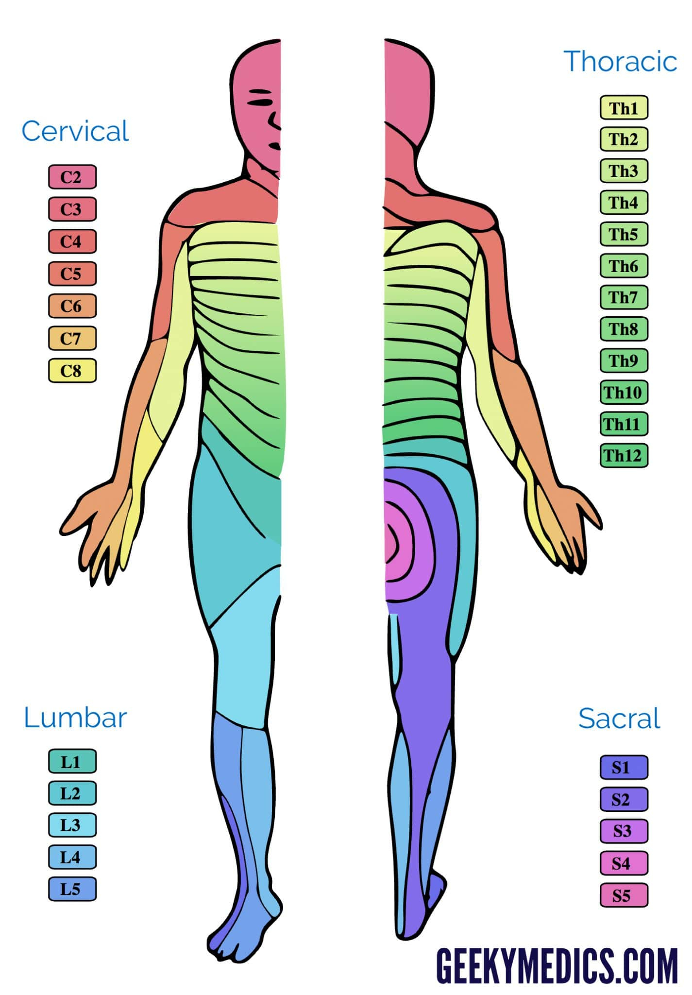
The dermatomes of the thoracic region are identified by letter-number combinations, which show how the region is connected in question and the sensory nerve that supplies the area. For example, the C1 spinal nerve does not have a dermatome, but those spinal nerves that are labeled C1-C8 and T9 is a reference with the belly button. Dermatomes are layered in horizontally on the trunk, those on the extremities tend to be in a longitudinal.
Dermatome Map
Dermatome maps are a common feature of textbooks that cover anatomy. However, the dermatome map is inconsistency both within and inter-textbook. Its name isn’t consistent, and some textbooks feature different maps on different pages. This is especially problematic when the authors of multiple chapters differ in their choice of dermatome map. Many textbooks use the maps of Foerster, Keegan, and Garrett however they don’t provide adequate references. Moreover, four textbooks use maps with no citations, and one of them is one that only cites secondary sources.
Dermatomes are the regions of skin that receives sensory input from the dorsal root of one spinal nerve. The dermatomes are not uniformly situated, but they tend to dip less inferiorly than horizontally. This is a natural variation and some tissues are covered by more than one dermatome. Additionally dorsal spinal nerve roots may have intrathecal intersegmental anastomoses to sensory neurons in Dorsal limbs.
L4-L5 Dermatome Map – Dermatome Map
Paediatric Neurological Examination OSCE Guide Geeky Medics