Herpes Zoster Dermatome Map – If you’ve ever wanted to know how the human dermatome chart will look, you’re in the right spot. Before we go to our map, we’ll look at what a dermatome is. What are the different kinds? And, most importantly, why is it essential to know about dermatomes in order to comprehend our body. Continue reading to learn more. You might be amazed! Here are some examples of dermatomes.
Gordelroos Herpes Zoster Huidarts
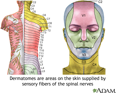
What is a Dermatome?
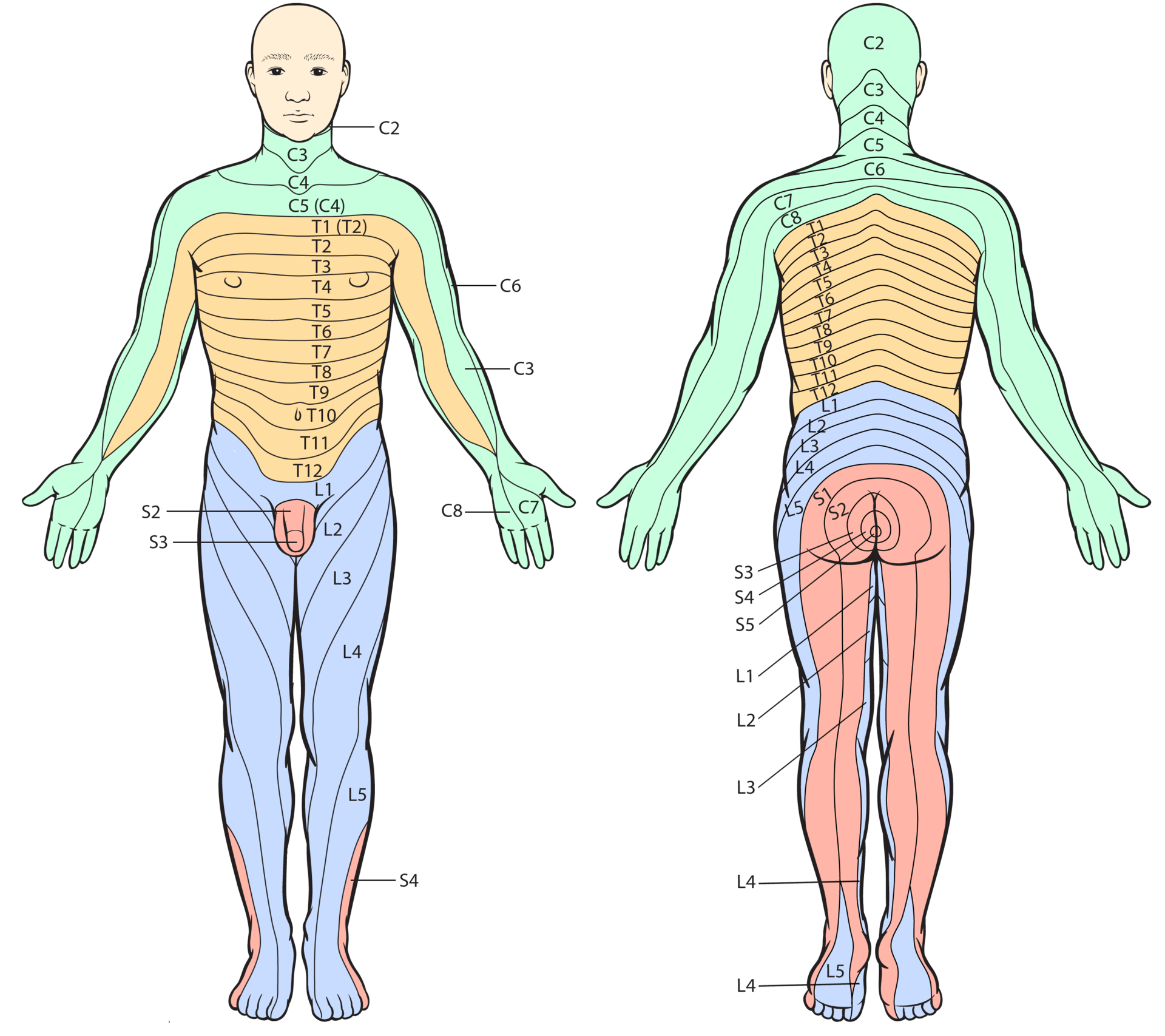
The term “dermatome” refers to a tissue that is a part of the spine. Dermatomes play a crucial role in allowing doctors to develop models of the cord, which help in diagnosing. Two maps are widely accepted by medical experts. They are the Keegan and Garret map and the Foerster map. These maps were created in the 1930s, and are frequently used. The trigeminal nerve as well as the maxillary nerve are the largest dermatomes.
Dermatomes are skin-like areas which are connected to a specific nerve. In the case of spinal cord injuries, pain may be experienced in a dermatome that is controlled by the nerve. Similar to the pain that is caused by an outbreak of shingles can be felt in specific spinal nerves. If you are experiencing nerve pain or neurological problem affecting the dermatome region, you need to see a doctor.
ALSO READ:[show-list showpost=5 category=”dermatome-map” sort=sort]
What are Some Examples of Dermatomes?
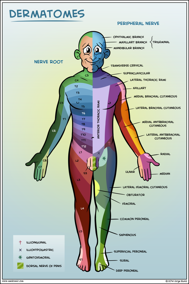
Dermatomes are the segments of skin that is supplied by one spinal nerve. These nerves provide motor, sensory, as well as autonomic information. They form part of the peripheral nerve system, that connects the brain to the rest of the body. A dermatome may suffer from a spinal cord injury. When one of these dermatomes becomes injured, it can be easily treated with the use of a local anesthetic.
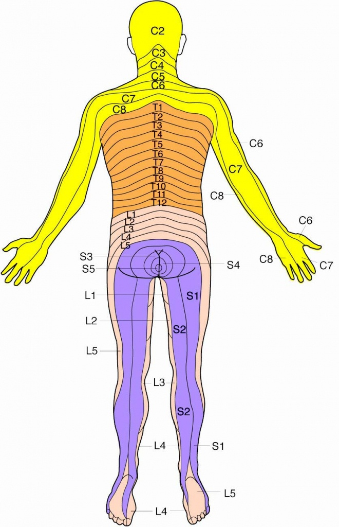
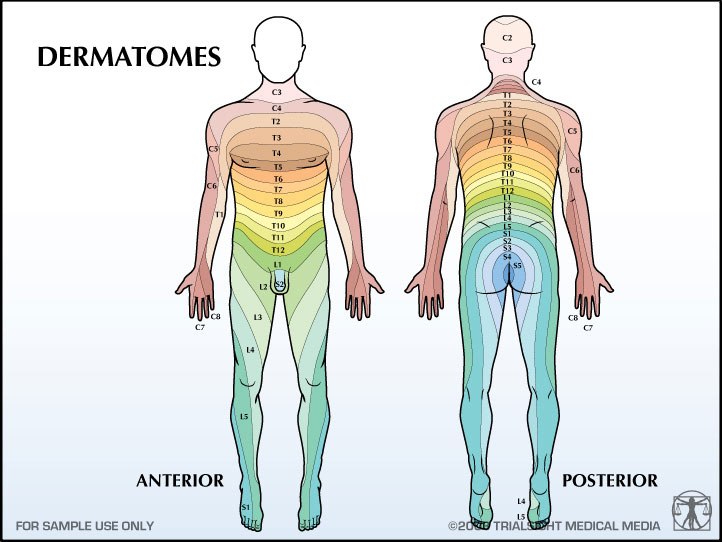
Dermatomes in the thoracic region are labeled with letters-numbers that illustrate the connection between the region in question and the sensory nerve that is responsible for this area. For instance, the C1 spinal nerve does not have a dermatome, but those spinal nerves that are labeled C1 – C8 T9, which corresponds to the belly button. Dermatomes are laid horizontally along the trunk, those on the extremities are typically long.
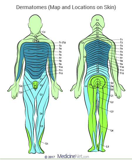
Dermatome Map
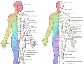
Dermatome maps are one of the common features of textbooks that teach anatomy. However, the dermatome maps is not consistent both within and inter-textbook. Its naming is inconsistent and certain textbooks have distinct maps on different pages. This can be particularly challenging in the event that the authors of various chapters differ in their choice of dermatome maps. Many textbooks use the maps of Foerster, Keegan, and Garrett however they don’t provide appropriate references. Furthermore, four textbooks make use of maps that do not have citations, such as one that refers to only secondary sources.
The dermatome is the area of the skin that receives sensory information from the dorsal root of a spinal nerve. The dermatomes are not uniformly located, but they tend to dip less inferiorly than horizontally. This is a normal variation and some tissues may be covered by multiple dermatomes. Also dorsal spinal nerve roots may have intrathecal intersegmental anastomoses to sensory neurons that originate from Dorsal limbs.
Herpes Zoster Dermatome Map – Dermatome Map
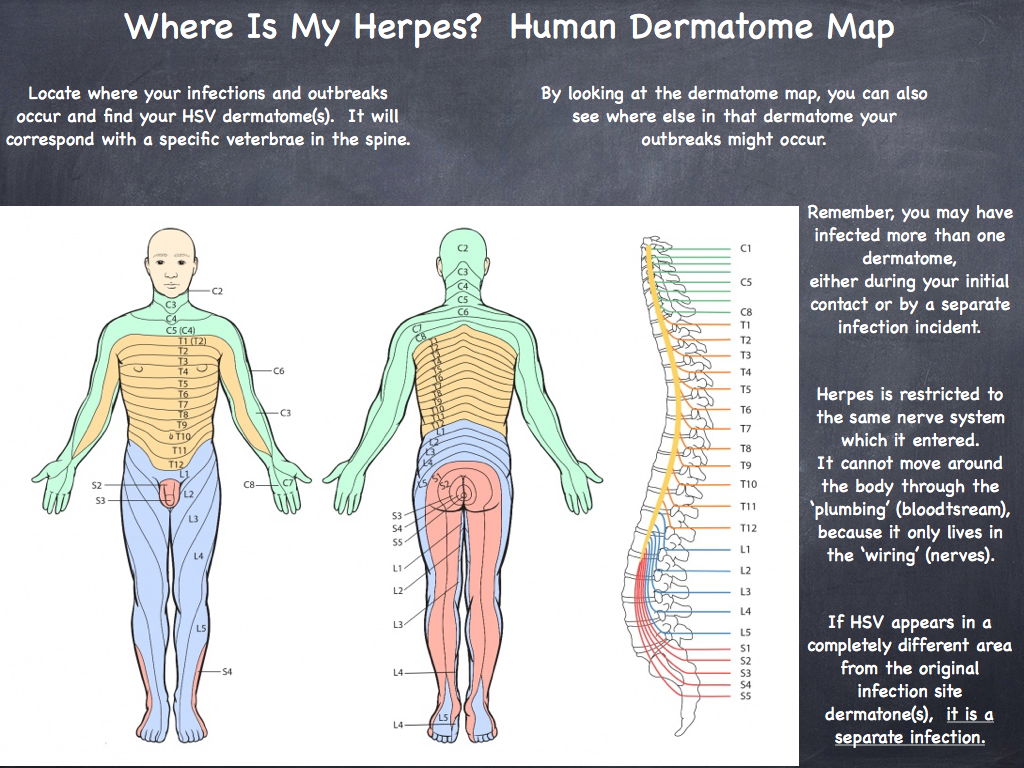
All About HSV Human Dermatome Map
Herpes Zoster Ophthalmicus Dermatome Chart Herpes Free Me

Dermatome Causes Symptoms Treatment Dermatome