Lower Body Dermatome Map – If you’ve ever wanted to know how the human dermatome map will look, you’re in the right spot. Before we get to an image, it’s important to look at what a dermatome is. What are the different kinds? The most important thing is why is it necessary to learn about dermatomes in order to understand the human body. Read on to find out more. You might be amazed! Here are some examples of dermatomes.
Pin On DERMATOMES
What is a Dermatome?
“dermatome,” or “dermatome” refers to a tissue that is a part of the spinal cord. Dermatomes can help physicians to build models of the cord, which are useful for diagnosis. Two major maps are accepted by medical specialists. These are: the Keegan and Garret map and the Foerster map. These maps were created in the 1930s and are still often employed. The trigeminal nerve , as well as the maxillary nerve are the largest dermatomes.
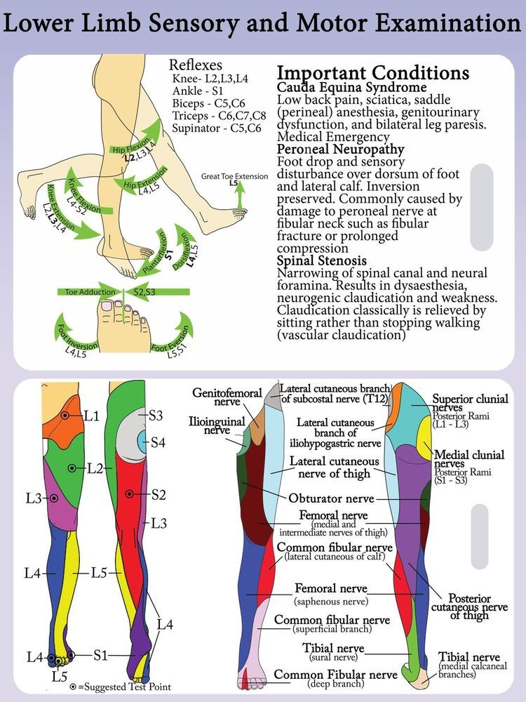
Dermatomes are skin-like areas that are attached to a particular nerve. In cases of spinal injury, the pain could be felt in a dermatome that is surrounded by the nerve. The same is true for the pain caused by shingles outbreaks is felt by specific spinal nerves. If you experience a discomfort or neurological issue involving the dermatome area, you must consult a physician.
ALSO READ:[show-list showpost=5 category=”dermatome-map” sort=sort]
What are Some Examples of Dermatomes?
A dermatome is a segment of skin that is provided by the spinal nerve. These nerves carry motor, sensory, and autonomic messages. They form an element of the peripheral nervous system, which connects the brain and all the body. A dermatome may get affected because of a spinal cord lesion. If one of these dermatomes gets injured, it is able to be easily treated with local anesthetic.
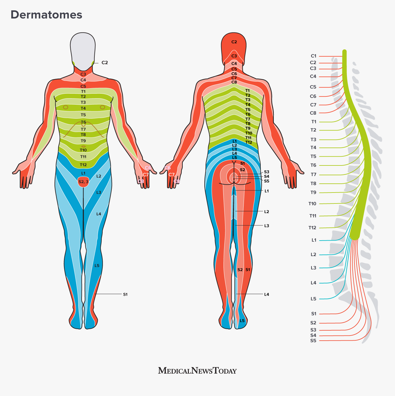
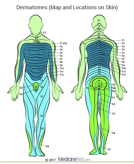
The dermatomes of the thoracic region are identified with letter-number combinations that show the connection between the area and the sensory nerve which supplies that area. For instance, the C1 spinal nerve does not have a dermatome. However, the other spinal nerves are identified as C1-C8 and T9 refers to belly button. Dermatomes are laid horizontally along the trunk, and dermatomes located on the extremities tend to be in a longitudinal.
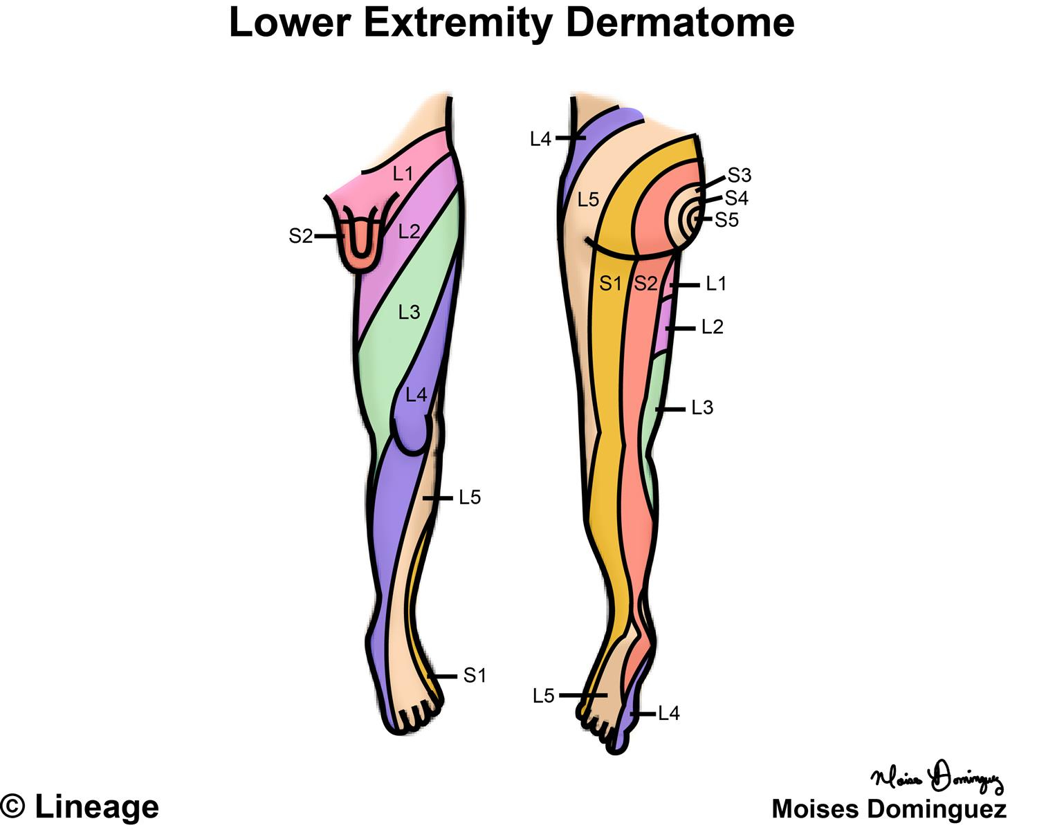
Dermatome Map
Dermatome maps are a common feature of textbooks that teach anatomy. The dermatome map is inconsistency both within and inter-textbook. Its name isn’t consistent and certain textbooks have different maps on various pages. This is especially problematic when the authors of different chapters differ in their choice of dermatome map. Most textbooks use Maps of Foerster, Keegan, and Garrett but don’t include appropriate references. Additionally, four textbooks employ maps that do not have citations, such as one that uses only secondary sources.
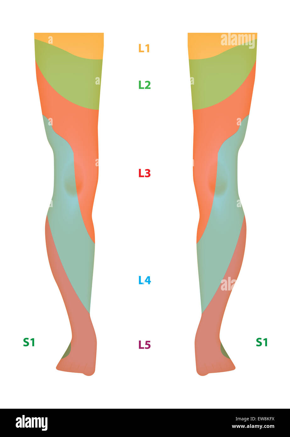
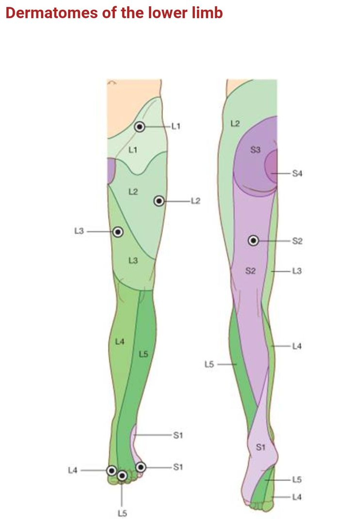
The dermatome is the area of the skin that receives sensory information from the dorsal branch of one spinal nerve. Dermatomes aren’t uniformly found, but they tend to dip lower than horizontally. This is a natural variation and certain tissues may be covered by multiple dermatomes. Furthermore, dorsal spinal rootlets may be anastomosed with intrathecal intersegmental sensory neurons in the dorsal limbs.
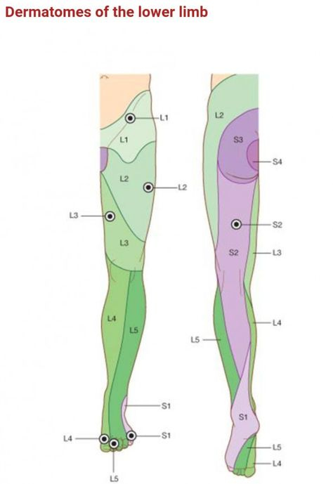
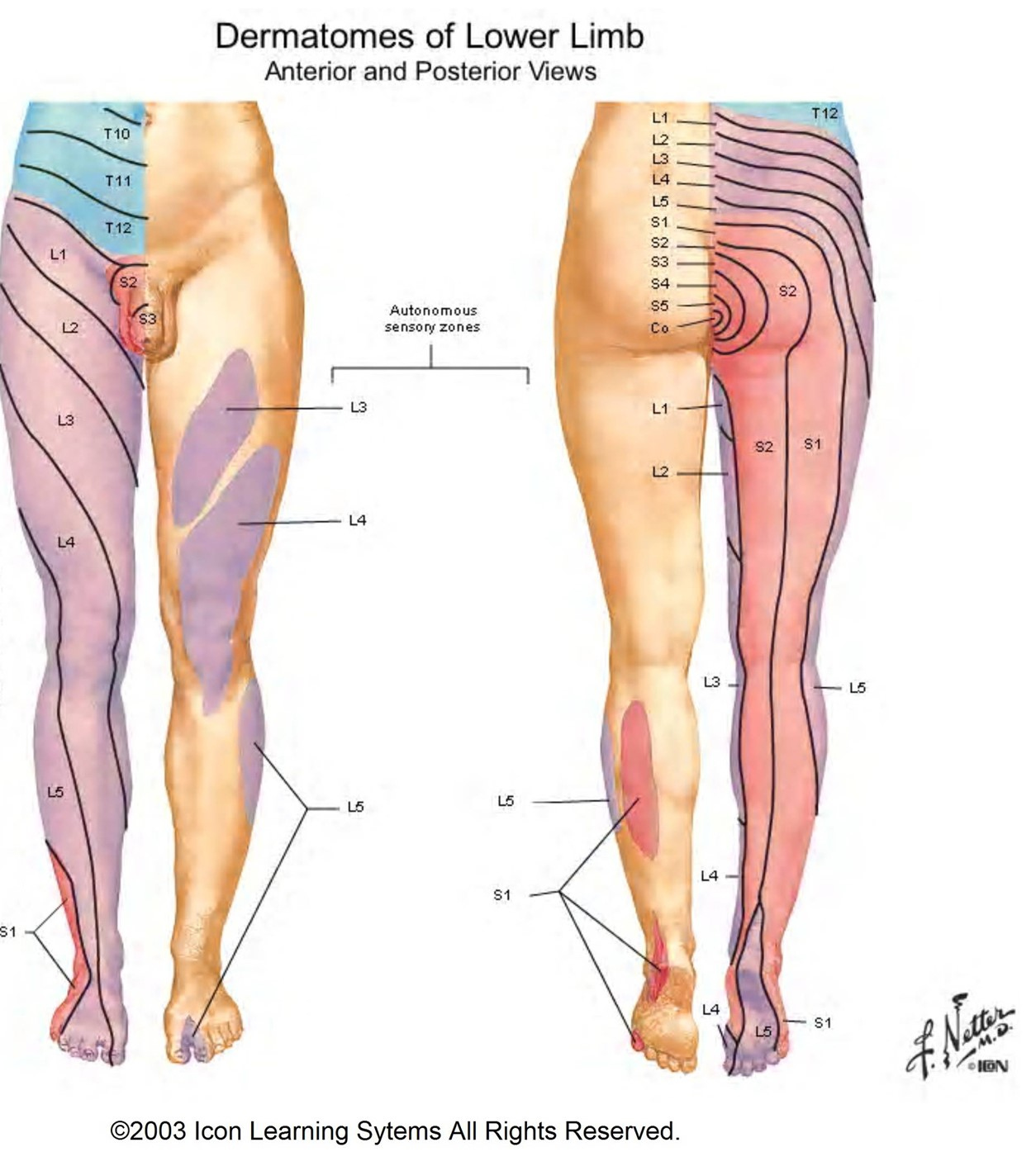
Lower Body Dermatome Map – Dermatome Map
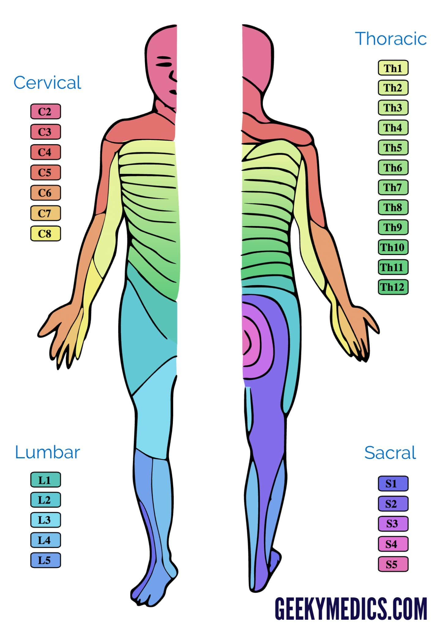
Dermatomes And Myotomes Sensation Anatomy Geeky Medics
Dermatomes And Myotomes
How Do Dermatomes Work Map Myotomes Vs Dermatomes Sensory Nerves